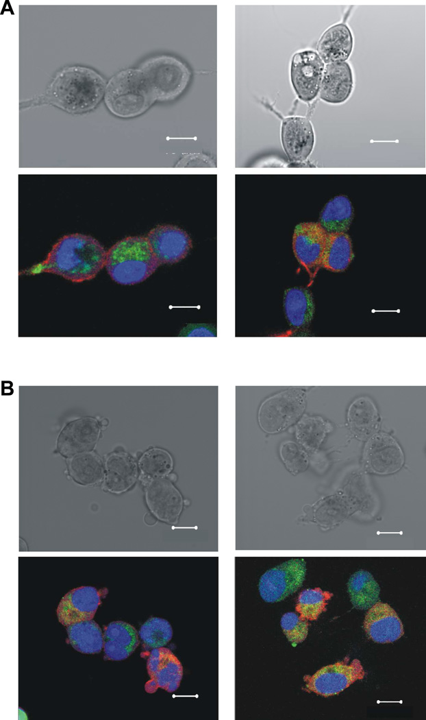
Figure 4.
Bright-field (Upper) and overlaid fluorescent (Lower) images of MCF7 (A) and MES-SA/Dx5 (B) cells after incubation with 25 nM of Fluorescein-PTX-DNA@AuNPs (Ex./Em. 494/521nm, green) for 6 h. Cellular Lights™ Actin-RFP (Ex./Em. 555/584nm, red, Invitrogen) was used for cytoplasmic actin staining, and DRAQ5 (Ex./Em 646/681nm, blue, Biostatus Ltd.) for nuclear staining. Scale bar is 10 µm.

