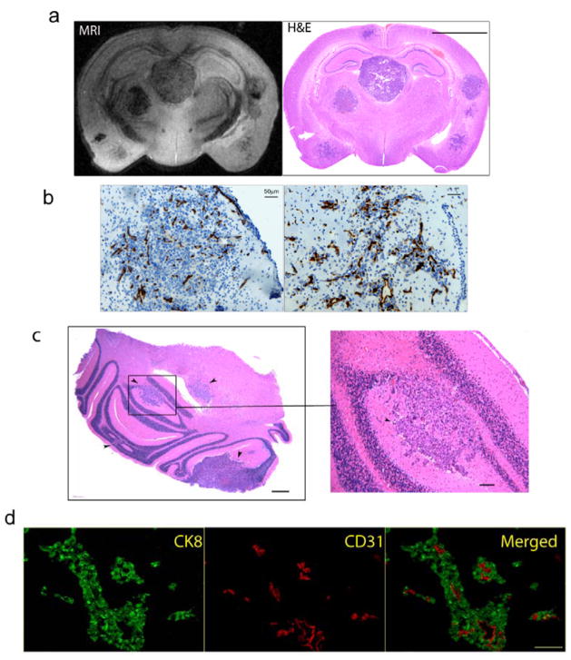
Figure 1. Histological characterization of the DU145/RasB1 brain metastasis model.
a. A high resolution coronal MRI section obtained on a fixed brain (left panel) and matching histological section (right panel) from an animal with multiple brain metastasis; (scale bar =2.5 mm). b. Microscopic images of tumors grow as an expanding nodular (left) and invasive (right) brain metastatic lesions; (scale bar =50 μm). Blood vessels were stained with CD31. c. Microscopic section of cerebellum with multiple metastases indicated by black arrows; (scale bar =200 μm; cropped section: scale bar =100 μm). d. Immuno-fluorescence staining of CK8 (green) and CD31 (red) on brain sections; (scale bar =50 μm).

