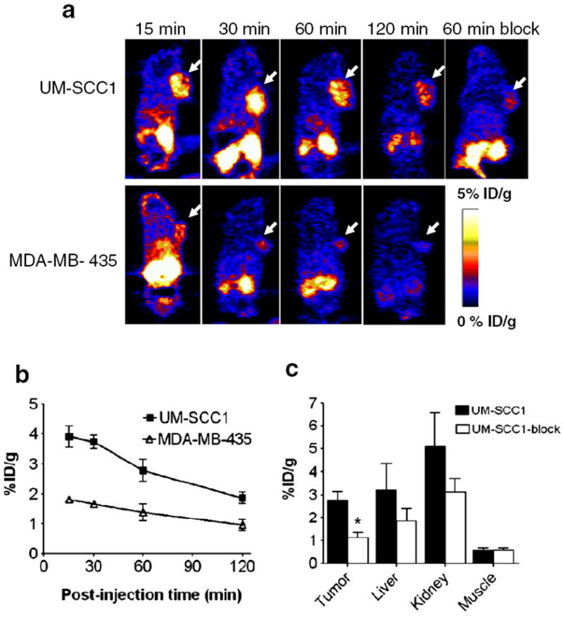
Fig. 6.

In vivo PET imaging of xenografted mice treated with 18F-FP-QQM. a Decay-corrected whole-body coronal microPET images of UM-SCC1 and MDA-MB-435 tumor–bearing mice at 15, 30, 60, and 120 min after injection of 3.7 MBq (100 μCi) of 18F-FP-QQM. The tumors are indicated by arrows. b ROI analysis of tumor uptake of 18F-FP-QQM at 15, 30, 60, and 120 min in UM-SCC1 and MDA-MB-435 tumor–bearing mice (n=6/group) as derived from static microPET images. c Quantification of 18F-FP-QQM uptake in UM-SCC1 tumor, liver, kidneys and muscle with and without the presence of a blocking dose of QQM peptide (n=6/group). Uptake values are shown as mean %ID/g±SD. *P<0.05
