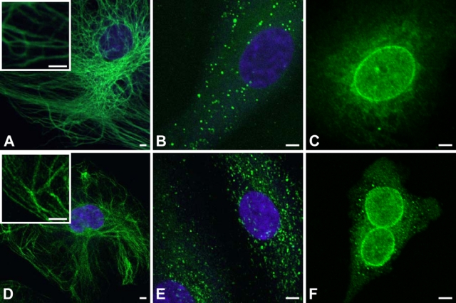
Figure 2.

Comparison of fluorescent polymerization-based amplification (FPBA) versus SA–Alexa 488 for immunofluorescent imaging of various antigens. (A) Human endothelial cells were immunostained with mouse anti-vimentin (1:50,000), biotinylated anti-mouse secondary (1:400), and fluorescently labeled by FPBA. (B) Human endothelial cells were stained for von Willebrand factor (vWF) with rabbit anti-vWF (1:250,000), biotinylated anti-rabbit secondary (1:500), and fluorescently labeled by FPBA. (C) Human endothelial cells were immunostained with mouse anti–nuclear pore complex protein (NPC; 1:1000), biotinylated anti-mouse secondary (1:400), and fluorescently labeled by FPBA. (D-F) Same as A through C, respectively, except that the fluorescent labeling was achieved using SA–Alexa 488 rather than FPBA. Scale bar is 5 µm. Images were taken on a confocal laser scanning microscope (CLSM) using a 40× oil objective for A, B, D, and E and a 63× oil objective for C and F. The nucleus is counterstained with 4′,6-diamidino-2-phenylindole dihydrochloride (DAPI; purple). DAPI staining in C and F confirmed that immunostaining is localized around the nucleus, but the DAPI stain is not depicted so as to more clearly indicate the NPC staining.
