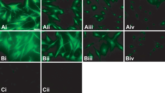
Figure 3.

Comparison of fluorescent polymerization-based amplification (FPBA) to other detection methods for the fluorescent imaging of nuclear pore complex proteins (NPCs) in human fibroblast cells. (A) FPBA is compared to (B) tyramide signal amplification (TSA) and (C) SA–Alexa 488. Cells were stained using (i) 1:102, (ii) 1:103, (iii) 1:104, or (iv) 1:105 dilution of primary antibody. In all cases, the secondary antibody was biotinylated anti-mouse at a 1:400 dilution. These images were obtained by epifluorescence microscopy, using a 20× objective lens, a 4-sec exposure time, and no binning of pixels. The scale bar is 200 µm. DAPI (4′,6-diamidino-2-phenylindole dihydrochloride) nuclear counterstaining verified that staining is centered on the nuclei, but the DAPI stain is not depicted so as to more clearly indicate the NPC staining.
