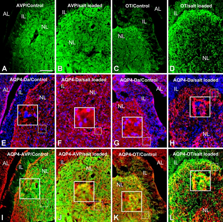
Figure 1.

Double immunohistochemical detection of arginine-vasopressin (AVP; green)/aquaporin-4 (AQP4; red) (A, B, E, F, I, J) and oxytocin (OT; green)/AQP4 (red) (C, D, G, H, K, L) in the mouse hypophysis before salt loading (A, E, I, C, G, K) and after 8 days of salt loading (B, F, J, D, H, L). The hypophysis is composed of three parts: the anterior lobe (AL), the intermediate lobe (IL), and the posterior or neural lobe (NL). In control mouse neurohypophysis, AQP4 immunolabeling colocalizes with cells (Hoëchst-stained nuclei) (E, G) and also with AVP- and OT-containing structures (I, K). No staining for AQP4 was detected in the intermediate lobe (E, G). AQP4 immunolabeling in the neural lobe (F, H), particularly in AVP- and OT-positive areas (J, L), was more intense after 8 days of salt loading than in controls. AQ, aquaporin 4; VP, vasopressin; Da, DAPI, 4,6-diamidino-2-phenylindole dihydrochloride. Scale bar: 100 µm.
