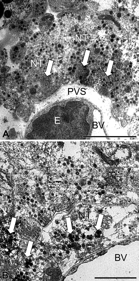
Figure 3.

Ultrathin section of the neurohypophysis showing aquaporin-4 (AQP4) immunolabeling in a blood vessel and its surroundings in control (A) and 8-day salt-loaded mice (B). In the control (A), the perivascular space is small with few nerve terminals containing labeled microvesicles, forming electron-dense clusters (arrow). In the salt-loaded mouse (B), the perivascular space is swollen and occupied by large nerve terminals rich in labeled neurosecretory granules and microvesicles (arrow). BV, blood vessel; E, endothelial cell; NT, nerve terminal; PVS, perivascular space. Scale bar: 2 µm (A); 1 µm (B).
