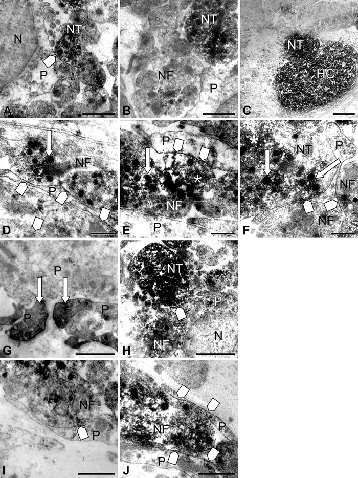
Figure 4.

High magnifications of immunoperoxidase staining in and around pituicytes in control (A-F) and 8-day salt-loaded mice (G-J). In the control, discontinuous labeling on pituicyte plasma membranes (square arrow, A) is observed; these labeled membrane segments were often in close contact with nerve terminals and fibers (square arrow, D-F), in which granules (arrow) and microvesicles (asterisk) were immunopositive. Neurosecretory granules and microvesicles contained in nerve terminals (B) and nerve fibers, including Herring cores, were labeled (C). In 8-day salt-loaded mice, immunolabeling for aquaporin-4 (AQP4) was observed on processes of pituicytes (G, arrow). AQP4 was detected along the plasma membrane of pituicytes, as in controls, but the staining was more intense (square arrow, H-J). N, nucleus; NT, nerve terminal; P, pituicytes; NF, nerve fiber; HC, Herring core. Scale bar: 1 µm (A-B, G-H); 2 µm (C); 0.5 µm (D-F, I-J).
