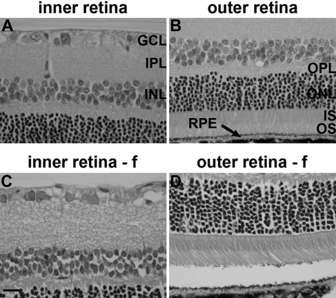
Figure 1.
Representative images of retinal histology after Davidson’s (A, B) and formalin (f; C, D) fixation, as delineated by hematoxylin and eosin staining. Scale bar = 15 µm. GCL, ganglion cell layer; IPL, inner plexiform layer; INL, inner nuclear layer; OPL, outer plexiform layer; ONL, outer nuclear layer; IS, inner segments of photoreceptors; OS, outer segments of photoreceptors; RPE, retinal pigment epithelium.

