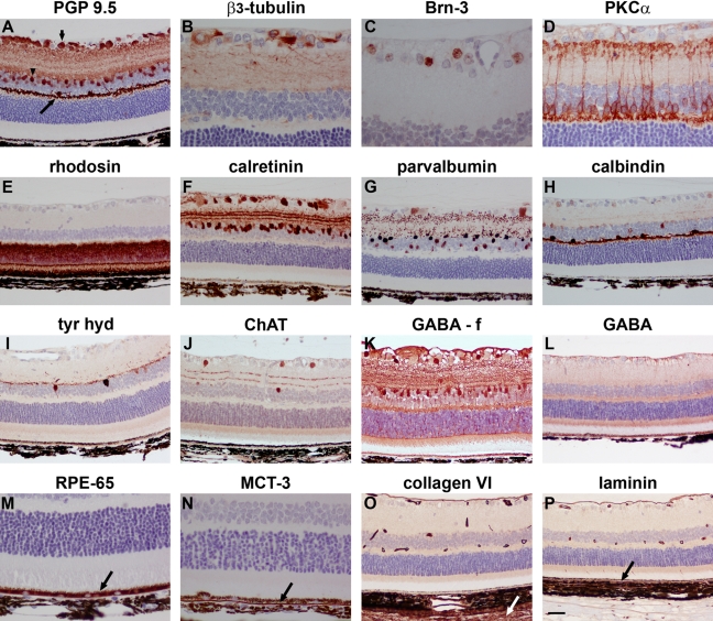
Figure 3.
Representative images of neuronal and non-neuronal markers in Davidson’s- and formalin- (f) fixed retinas, as shown by immunohistochemistry. (A) Retinal ganglion cells (RGCs) (short arrow), amacrine cells (arrowhead), and horizontal cells (long arrow) labeled by the pan-neuronal marker PGP 9.5. (B) RGC somata, dendrites, and axons labeled by β3-tubulin. (C) RGC nuclei labeled by Brn-3. (D) Bipolar cells and their processes terminating in the inner and outer plexiform layers labeled by protein kinase C (PKC)-α. (E) Photoreceptor cell bodies and their segments labeled by rhodopsin. (F) Amacrine cells and RGCs labeled by calretinin with 3 layers of terminals visible in the inner plexiform layer (IPL). (G) RGCs, amacrine cells, and their terminals labeled by parvalbumin. (H) Horizontal cells and their processes labeled by calbindin. (I) Putative dopaminergic amacrine cells as labeled by tyrosine hydroxylase (tyr hyd). (J) Putative cholinergic amacrine cells and 2 layers of terminals visible in the IPL as labeled by choline acetyl transferase (ChAT). (K) GABAergic amacrine cells as labeled by GABA in a formalin-fixed retina. (L) Lack of GABA immunolabeling in a Davidson’s-fixed retina. (M) Retinal pigment epithelium (RPE) labeled by RPE-65. (N) Basolateral surface of RPE labeled by MCT3. (O) Labeling of inner limiting membrane, blood vessels, and sclera (arrow) by collagen VI. (P) Labeling of inner and outer (arrow) limiting membrane and blood vessels by laminin. Scale bar: B-D, M, N = 15 µm; A, E-L, O, P = 30 µm.

