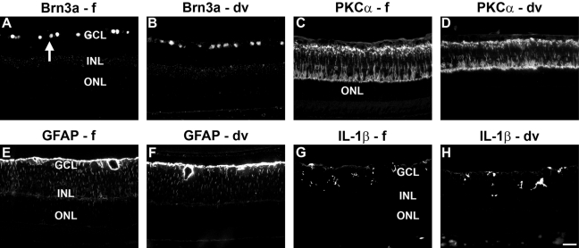
Figure 6.
Representative images of neuronal and non-neuronal markers in formalin-fixed (f) and Davidson’s-fixed (dv) retinas, as shown by fluorescent immunohistochemistry. Retinal ganglion cell (RGC) nuclei (arrow) labeled by Brn-3a (A, B) and bipolar cells and their processes labeled by protein kinase C (PKC)-α (C, D) in normal retinas. Astrocytes and Müller cells labeled by glial fibrillary acidic protein (GFAP) (E, F) and microglia labeled by interleukin (IL)-1β (G, H) in retinas of lipopolysaccharide (LPS)-injected eyes. Scale bar = 30 µm. GCL, ganglion cell layer; INL, inner nuclear layer; ONL, outer nuclear layer.

