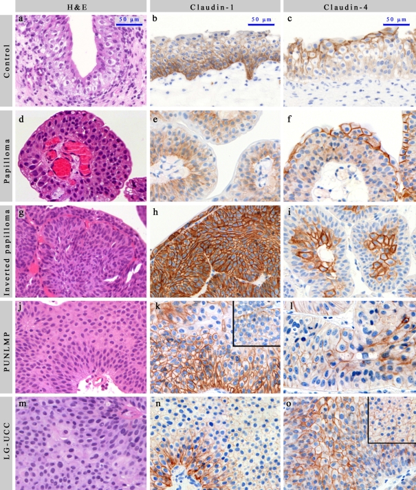
Figure 1.
Each photograph (including inserts) was taken with standard adjustments. Scale bar: 50 µm. (A, D, G, J, M) H&E in normal, UP, IUP, PUNLMP, and LG-UCC, respectively. (B, E, H, K, N) Claudin-1 in normal, UP, IUP, PUNLMP, and LG-UCC, respectively. (C, F, I, L, O) Claudin-4 in normal, UP, IUP, PUNLMP, and LG-UCC, respectively. LG-UCC expressing claudin-4 over the median (O) and under the median (insert of O). PUNLMP expressing claudin-1 over the median (K) and under the median (insert of K). UP = urothelial papilloma; IUP = inverted urothelial papilloma; PUNLMP = papillary urothelial neoplasm of low malignant potential; LG-UCC = low-grade urothelial cell carcinoma. Only linear adjustments of brightness/contrast and color balance were applied for whole images in order to create uniform-looking pictures for the composite image.

