
Figure 1.
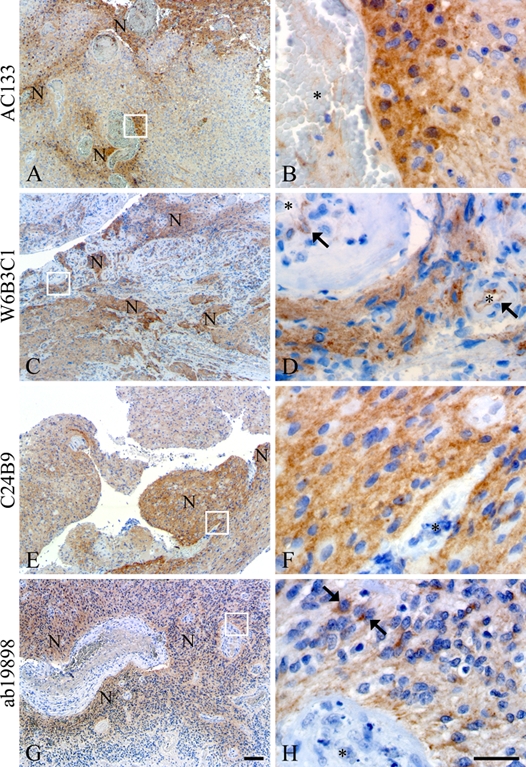
All clones AC133 (A, B), W6B3C1 (C, D), C24B9 (E, F), and ab19898 (G, H) identified niches of perivascular tumor cells in the glioblastomas. W6B3C1 also showed staining of the basal endothelial membrane of most blood vessels (arrows, D). Clone ab19898 intensely stained dispersed cells within the niches (arrows, H). Vessel lumen is indicated by an asterisk (B, D, F, H), and N denotes niches. Scale bars: 50 µm (A, C, E, G); 30 µm (B, D, F, H).