
Figure 9.
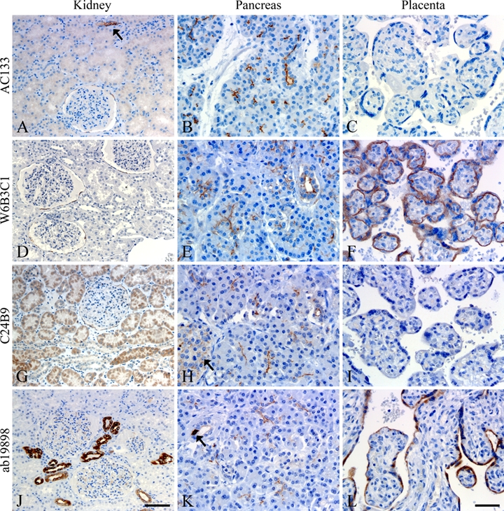
Comparative staining of kidney (A, D, G, J), pancreas (B, E, H, K), and placenta (C, F, I, L) tissues. A varying degree of CD133 positivity was observed in kidney glomeruli, tubuli, and collecting ducts between the clones (A, D, G, J). Arrow in A shows a stained collecting duct. Positive reaction in pancreas was mainly constricted to the apical/endoluminal surface of glandular epithelial and ductal cells (B, E, H, K), but some diffuse staining of islets of Langerhans (arrow, H) and cytoplasmatic staining of pancreatic ductal cells could be observed (arrow, K). Placental tissue was negative using AC133 and C24B9 (C, I) and positive in trophoblasts/syncytiotrophoblasts of chorionic villi using W6B3C1 and ab19898 (F, L). Scale bars: 100 µm (A, D, G, J); 50 µm (B, C, E, F, H, I, K, L).