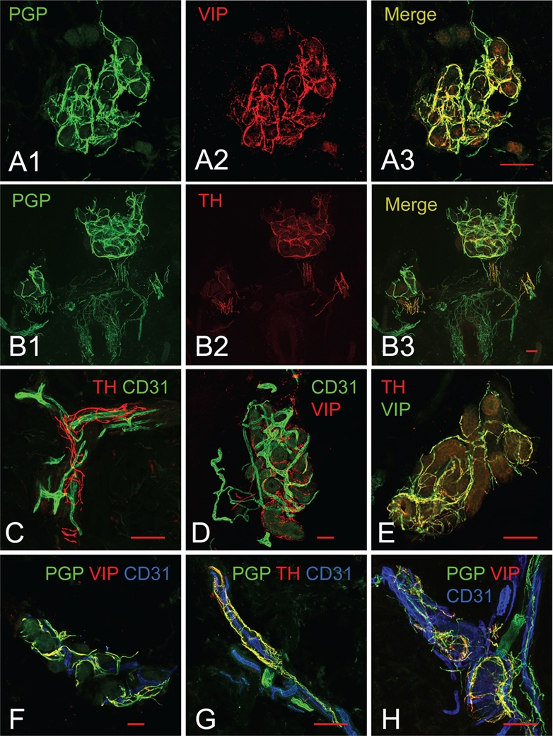
Figure 2.

Examples of images obtained through immunostaining protocols. (A, B) Double staining with primary antibodies raised from the same species (rabbit). (A1) Rabbit anti-PGP9.5, stained with the conventional method, reveals all nerve fibers surrounding sweat glands. (A2) Rabbit anti-VIP, amplified by the tyramide signal amplification system (TSA), selectively highlights the sympathetic cholinergic fibers surrounding the sweat glands. (A3) Co-localization of VIP-ir fibers with PGP9.5-ir fibers is observed surrounding sweat glands. (B1) Rabbit anti-PGP9.5, stained with the conventional method, reveals all nerve fibers surrounding sweat glands, hair follicles, and arrector pili muscles. (B2) Rabbit anti-TH, amplified by TSA, selectively highlights the sympathetic adrenergic fibers in the arrector pili muscles and some of the sweat gland tubules. (B3) Co-localization of TH-ir fibers with PGP9.5-ir fibers is observed surrounding sweat glands and arrector pili muscles but not within the sensory fibers surrounding the hair follicle. (C, D) Double staining with parallel signal amplification systems of two weakly expressed antigens. (C) Mouse anti-CD31 is amplified by streptavidin-biotin-fluorochrome (sABC) and rabbit anti-TH is amplified by TSA, thus highlighting the sympathetic adrenergic innervation of the cutaneous vasculature. (D) Mouse anti-CD31 is amplified by sABC and rabbit anti-VIP is amplified by TSA, highlighting the complicated vasculature and cholinergic innervation of sweat glands. (E) Parallel amplification of rabbit anti-VIP and rabbit anti-TH shown with co-localized sympathetic adrenergic and sympathetic cholinergic fibers. (F–H) Parallel amplification of rabbit anti-TH (or -VIP) and mouse anti-CD31 for staining of nerve fibers and blood vessels with the conventional method of nerve fibers using rabbit anti-PGP9.5. (F) The total innervation and subpopulation of sympathetic adrenergic nerves around a sweat gland with the accompanying vasculature. (G) The total innervation and subpopulation of sympathetic adrenergic nerves surrounding a dermal blood vessel. (H) The total innervation and subpopulation of sympathetic cholinergic nerves around a sweat gland with the accompanying vasculature. Scale bars indicate 100 µm. AP, arrector pili muscles; BV, blood vessels; HF, hair follicles; SG, sweat glands.
