
Figure 6.
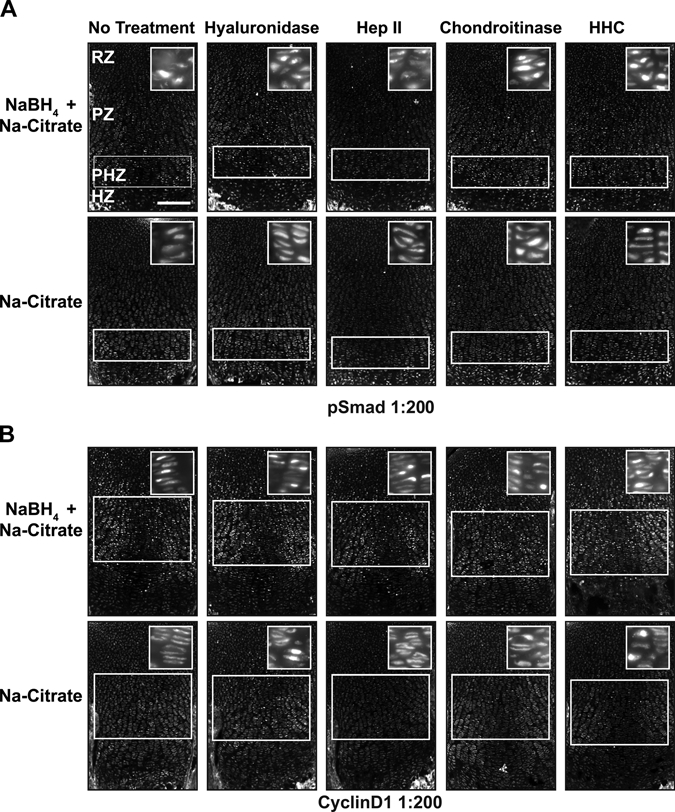
Combining pretreatments optimizes signal detection. Immunofluorescence detection of the stated antigens was performed on sections of P0 mouse growth plate cartilage subjected to the designated combinations of pretreatments (header and left of rows). Antigen retrieval (Na-citrate) alone results in a high level of cytosolic signal for anti-pSmad (A) and anti-cyclinD1 (B), as demonstrated by the clearly discernable structure of the columnar chondrocytes of the proliferative zone. (A) Detection of pSmad in the prehypertrophic zone (PHZ) is improved with Na-citrate pretreatment in combination with NaBH4 (note loss of visible columns). Hyaluronidase, chondroitinase, or a combination of three sugar-degrading enzymes (hyaluronidase + heparinase II + chondroitinase [HHC]) reveals nuclear staining in the proliferative zone (PZ) and resting zone (RZ) when used in combination with Na-citrate and NaBH4. (B) Similarly, NaBH4 and Na-citrate pretreatment significantly increased the signal:noise ratio for cyclinD1 in the PZ and RZ. The addition of hyaluronidase and chondroitinase or HHC further increased the signal and the specific localization of the antigen. Multiple pretreatments reveal higher levels of signal in the proliferative chondrocytes relative to the resting chondrocytes. White boxes indicate location where signal is present or expected. Scale bar in A–C, 200 µm.