
Figure 7.
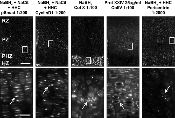
Optimal conditions reveal proper localization of antibodies. For each pretreatment condition (header), a low-magnification immunofluorescence image (top) and a high-magnification image (bottom) corresponding to the white box (top) are presented. (A) Optimal conditions for pSmad, cyclinD1, and pericentrin include NaBH4, Na-citrate, and hyaluronidase + heparinase II + chondroitinase (HHC). High-magnification images of boxed regions show proper localization of antibodies (white arrows). pSmad is nuclear localized through the growth plate. CyclinD1 is nuclear localized in dividing cells in the resting zone (RZ) and proliferative zone (PZ). Pericentrin is found throughout the growth plate at the base of primary cilia. The optimal condition for the matrix protein collagen X (Col X) is NaBH4 alone, but for collagen IV (Col IV), treatment with proteinase XXIV (Prot XXIV) produces optimal results. Col X is found in the matrix of the HZ, whereas Col IV is found in the matrix surrounding resting and proliferative chondrocytes (RZ and PZ). Scale bars: in A, 200 µm for 10× images and 25 µm for 40× images.