
Figure 8.
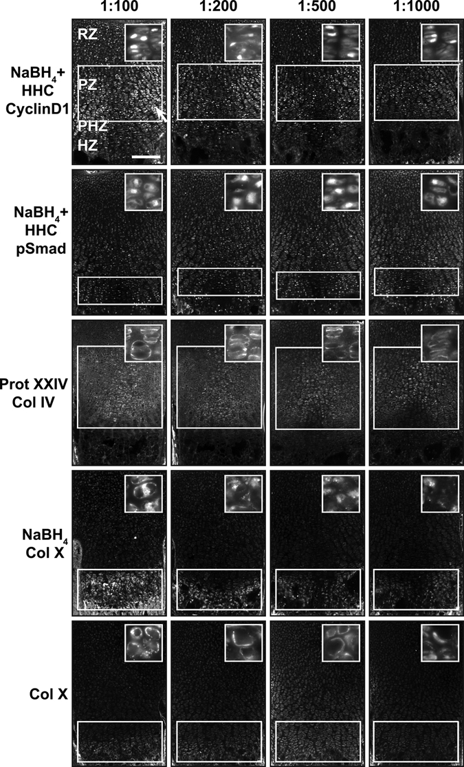
Antibody dilution can be increased with cartilage pretreatment. (A) Optimal pretreatment conditions of cyclinD1, pSmad, collagen IV (Col IV), and collagen X (Col X) allow visualization of antibodies at lower dilutions, up to 1:1000. CyclinD1 has nonspecific cytoplasmic staining (white arrow) when the concentration is too high (1:100). Note how the contrast between signal in the resting and the proliferative zone increases with antibody dilution. Optimal conditions increase the sensitivity of immunofluorescence for Col X. If there is no pretreatment, Col X has only weak signal at a dilution of 1:100. However, the signal:noise ratio is much higher for Col X at 1:100 when it is pretreated with NaBH4, and signal can be visualized even at a dilution of 1:1000. White bars indicate highest levels of expression. White boxes indicate location where signal is present or expected. Scale bar: in A, 200 µm.