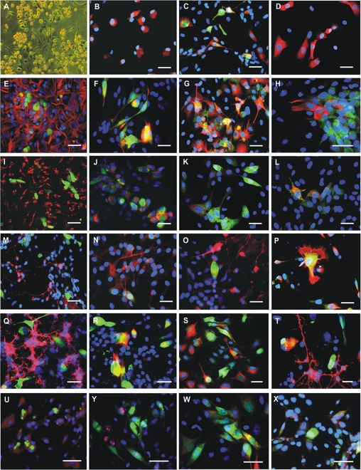
Figure 1.
Human umbilical cord blood neural-like stem cells (HUCB-NSCs) in a culture (A–D), and neural marker expression in HUCB-NSCs co-cultured with neonatal rat brain cells: astrocytes (E–H), microglia (I–L), neurons (M–P), oligodendrocytes (Q–T), and endothelial cells (U–Y). Specific immunostaining for nestin (B), NF200 and TUJ1 (C), or S-100β (D) in the HUCB-NSC population. Cells stained with Texas red after immune reactions were detected simultaneously, and Hoechst 33258 staining showed their nuclei (blue). Neonatal rat astrocyte primary culture monolayer immune labeled with anti–glial fibrillary acidic protein (GFAP) antibody (red) co-cultured with HUCB-NSCs (green by CMFDA) (E). Neural differentiation of HUCB-NSCs cultured with rat astrocytes. HUCB-NSC (green by CMFDA and red by Texas red after phenotype-specific immune reaction) can be detected in the presence of all cells forming astrocyte primary culture shown after using Hoechst 33258 (blue). Co-localization of red and green labeling appears yellow after overlaying these two images. NF200 (F) and TUJ1 (G) expressing cells display neuron-like morphology with axonal projection. S-100β expression (red) is detected yellow in some green prelabeled HUCB-NSC (H). Microglia of rat primary culture stained with anti-ED1 antibody (red) co-cultured with HUCB-NSCs (green by CMFDA) (I). Immunophenotyping of NF200 (J), TUJ1 (K), and S-100β (L) positive cells (red) in HUCB-NSCs (green prelabeled with CMFDA) co-cultured with rat microglia. Neonatal rat postmitotic neuron primary culture monolayer immunostained with anti-TUY1 antibody (red) co-cultured with HUCB-NSCs (green by CMFDA) (M). Immunolabeling for NF200 (N), TUJ1 (O), and O4 (P) positive cells (red) in HUCB-NSCs (green prelabeled with CMFDA) cultured in the presence of mature rat neurons. Oligodendrocyte-enriched rat primary culture stained with anti-NG2 antibody (red) co-cultured with HUCB-NSCs (green by CMFDA) (Q). Immune reaction depicting NF200 (R), TUJ1 (S), and GalC (T) positive cells (red) among HUCB-NSCs (green prelabeled CMFDA) co-cultured with rat oligodendrocytes. Endothelial cells (t-END line) monolayer stained with anti-vWF antibody (red) co-cultured with HUCB-NSCs (green by CMFDA) (U). Immunostaining for Ki67 (Y), TUJ1 (W), and S-100β (X) positive cells (red) in HUCB-NSC population (green prelabeled with CMFDA) cultured in the presence of t-END cells. Scale bars = 20 µm.

