
Figure 2.
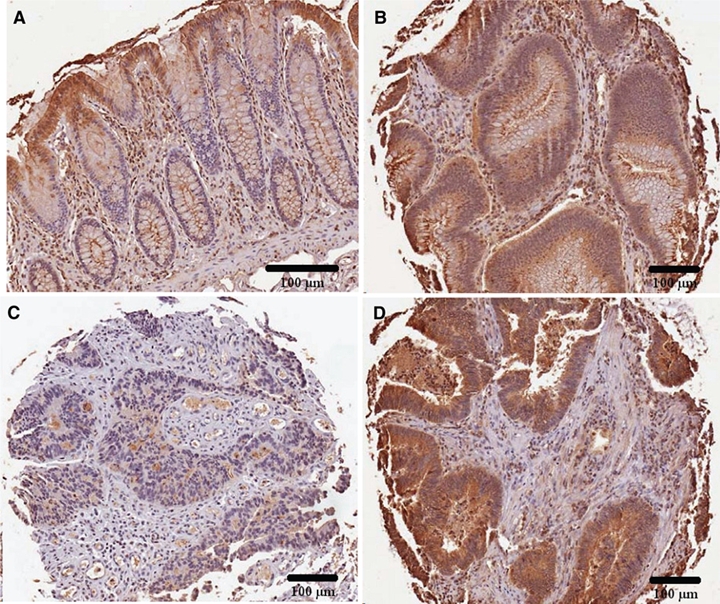
Overall pattern of arylsulfatase B (ARSB) staining in normal, villous adenomas, and adenocarcinomas. (A) Normal tissue demonstrates a distinct pattern of ARSB staining, with increased intensity at the luminal surface where differentiated cells are present. Along the mid-portion of the crypts, ARSB staining is absent, except for an occasional positively stained cell. At the base of the crypts, rare ARSB-positive cells are present. (B) In the villous adenomas, the distinctive topography of ARSB staining seen in the normal crypt is absent, and there is uniform epithelial staining. (C, D) These adenocarcinomas demonstrate loss of the distinctive overall pattern of ARSB staining seen in the normal tissue, with low and high percentages of cytoplasmic staining and intensity. ARSB immunostaining is brown; hematoxylin counterstain is blue. Scale bar is 100 µm.