
Figure 4.
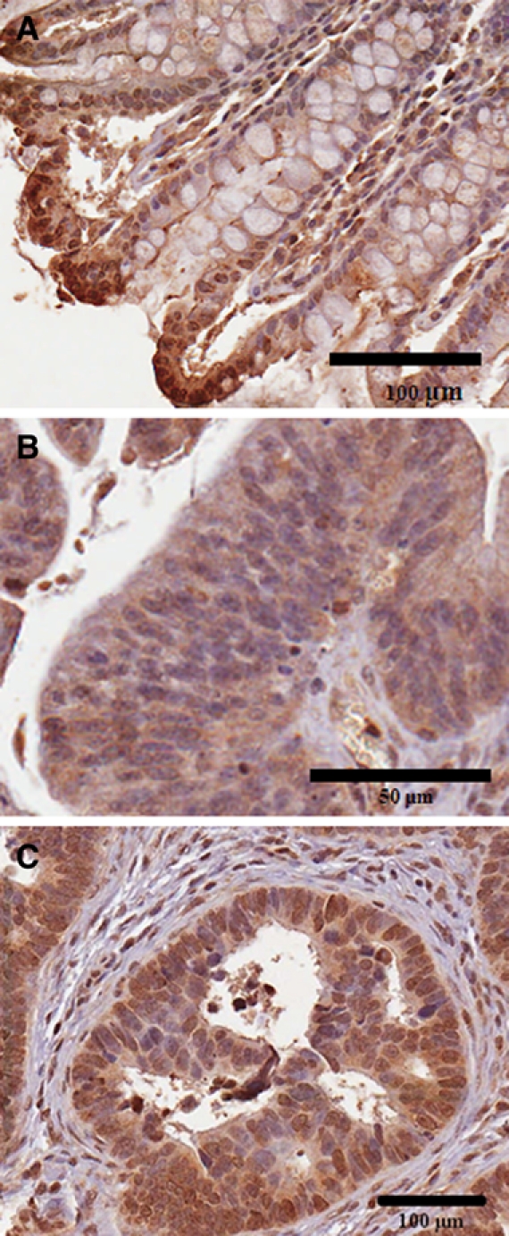
Differences in epithelial nuclear staining between normal and malignant tissue. (A) In the normal epithelial cells, nuclear staining is present in some of the cells at the luminal border and is intense. Nuclear membrane staining is also prominent in some of the cells. Scale bar is 100 µm. (B) In the villous adenoma, the nuclear staining is generally negative but with occasional positive cells. Nuclear membrane staining is prominent in some of the cells. Scale bar is 50 µm. (C) In the malignant tissue, there is variation in the extent of nuclear staining, similar to the variation in cytoplasmic staining. In some of the adenocarcinomas, nuclear staining is predominantly negative, whereas in other cores, many of the nuclei and nuclear membranes appear positive. ARSB immunostaining is brown; hematoxylin counterstain is blue. Scale bar is 100 µm.