
Figure 6.
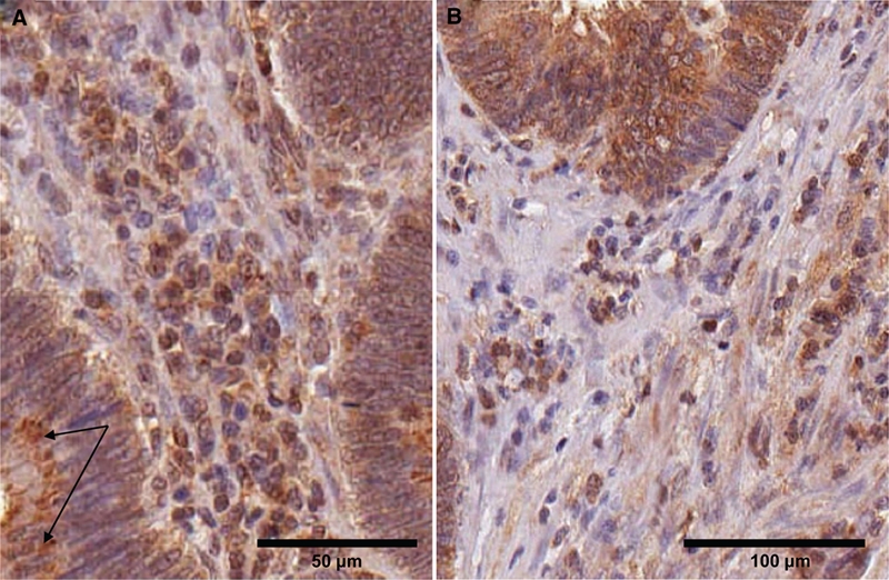
Variation in arylsulfatase B (ARSB) staining in the stroma and cytoplasmic inclusions positive for ARSB cells. (A) In the villous adenomas, the stromal cells appear more homogeneous than in the normal cores (Fig. 4A and Fig. 5). Distinct ARSB-positive inclusions are seen in the cytoplasm of the epithelial cells; arrows indicate representative inclusions. Scale bar is 50 µm. (B) In the adenocarcinomas, more variability is apparent in the stromal cells. Many are ARSB positive, with an increase in positively staining elongated cells that may be myofibroblasts. ARSB immunostaining is brown; hematoxylin counterstain is blue. Scale bar is 100 µm.