
Figure 1.
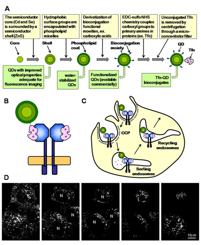
(A) Generation of transferrin (Tfn)–quantum dot (QD) bioconjugates. (B) Two molecules of Tfn (iron bound; asterisks) bind each transferrin receptor (TFR) dimer at the plasma membrane. In this particular example, one QD–Tfn and one unlabeled Tfn bind a TFR dimer. (C) TFR–Tfn complexes are internalized via clathrin-coated pits (CCP) and delivered to endosomes by clathrin-coated vesicles. Upon endosome acidification, iron is released from Tfn, and then the TFR–Tfn complexes are recycled back to the plasma membrane (PM) via recycling endosomes. (D) Endocytic uptake of Tfn–QD580 leads to a tubulo-vesicular staining throughout the cell. Confocal images were collected via a vertical z-scan with a 1-µm interval.