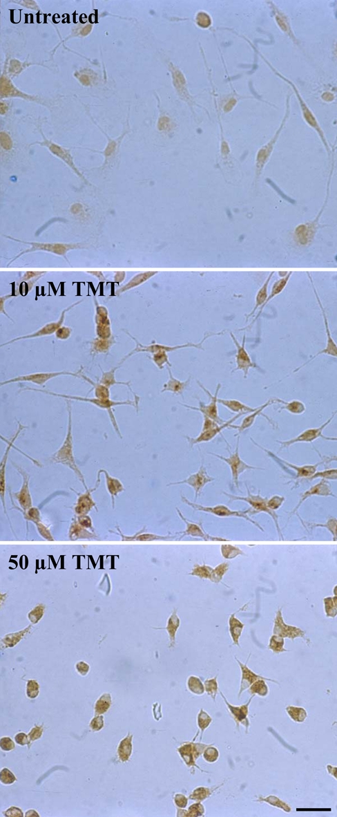
Figure 5.

Protease-activated receptor–1 (PAR-1) immunostaining in rat primary microglia. Untreated microglial cells show a weak diffuse staining for PAR-1. After exposure to 10 µM trimethyltin (TMT) for 24 hr, a stronger immunoreactivity for PAR-1 is evident in activated microglial cells, which show a rounding up of the cell body and a retraction of cell processes. Following exposure to 50 µM TMT, almost all surviving microglial cells are rounded and intensely positive for PAR-1. Photographs show a representative image for five independent experiments. (Scale bar = 20 µm)
