Figure 2.

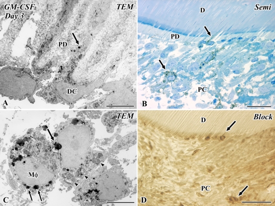
Electron micrographs (A, C) and a semithin section (B), as well as an Epon block after the cutting of ultrathin sections (D) of granulocyte macrophage colony-stimulating factor (GM-CSF) immunoreactivity in the transplanted tooth at 3 days after operation. D, dentin; PC, pulp chamber; PD, predentin; TEM, transmission electron microscope. (A–D) Secretory granule- or lysosome-like structures in the Golgi area are positive for GM-CSF in the macrophages (Mφ) and dendritic cells (DC) (arrows in A and C). Some of these positive cells are arranged along the pulp-dentin border and extend their cellular processes into the dentinal tubules. These cells possess peculiar cell organelles such as multivesticular bodies and tubulovesicular structures, and macrophages contain phagosomes (arrowheads) in addition to these cell organelles (A, C). Bars: D = 50 µm; B = 25 µm; A, C = 5 µm.
