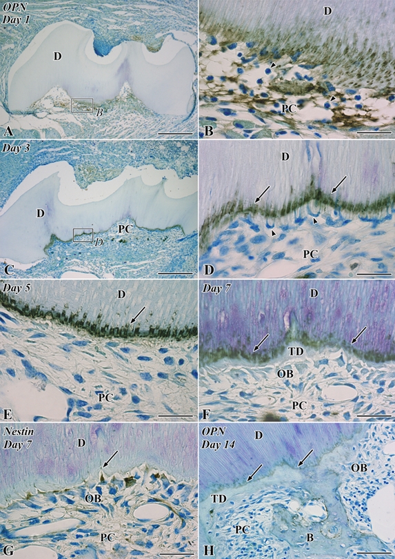
Figure 3.

Osteopontin (OPN; A–F, H) and nestin immunoreactivity (G) in the transplanted teeth at 1 (A, B), 3 (C, D), 5 (E), 7 (F, G), and 14 (H) days after operation. B, bone; D, dentin; OB, odontoblast-like cell; PC, pulp chamber; TD, tertiary dentin. (A) Intense OPN-positive reactions are observed throughout the pulp chamber. (B) Higher magnification of the boxed area labeled by B in A. Numerous neutrophils are recognized through the pulp chamber (arrowheads), and OPN-positive reactions are densely distributed in the fibrin networks and the pulp-dentin border, including the dentin matrix. (C) OPN immunoreactions are localized in the pulp-dentin border and certain pulpal cells. (D) Higher magnification of the boxed area labeled by D in C. The mineralization front of the preexisting dentin represents continuous intense OPN-positive reactions (arrows), and the cells with an irregular shape appear along the pulp-dentin border and extend their cellular processes into the dentinal tubules (arrowheads). (E) The mineralization front of the preexisting dentin maintains intense OPN-positive reactions (arrow), and the irregular-shaped cells disappear from the pulp-dentin border. (F) The continuous OPN immunoreactions are observed at the boundary between the preexisting dentin and tertiary dentin (arrows). (G) Nestin-positive odontoblast-like cells are arranged along the pulp-dentin border (arrow). (H) The continuous OPN immunoreactions are recognized both at the boundary between the pre- and postoperative dentin (arrows) and around the bone matrix. Bars: A, C = 250 µm; H = 50 µm; B, D, E–G = 25 µm.
