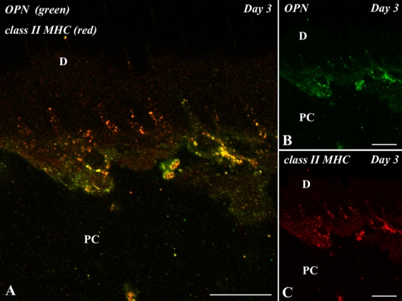
Figure 6.

Double immunofluorescent section with anti–osteopontin (OPN) (green) and class II major histocompatibility complex (MHC) (red) antibodies of the pulp-dentin border of the transplanted tooth at 3 days after operation. D, dentin; PC, pulp chamber. (A–C) Class II MHC-positive cells with dendritic features appearing along the pulp-dentin border represent a positive reaction for OPN. Bars = 10 µm.
