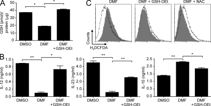
Figure 2.
DMF induces mouse type II macrophages and type II DCs. (A) BMDCs were incubated with DMSO, 70 µM DMF, or DMF + 1 mM GSH-OEt, and GSH content was determined by a colorimetric assay (mean ± SEM; *, P < 0.001). (B) DCs were treated with DMSO, 70 µM DMF, or DMF + 1 mM GSH-OEt and then stimulated with LPS for 18 h. Culture supernatants were harvested, and the indicated cytokines were determined by ELISA (mean ± SEM; *, P < 0.01; **, P < 0.001). (C) DCs were incubated with DMSO, 70 µM DMF, DMF + 1 mM GSH-OEt, or DMF and 1 mM NAC for 2–4 h, and intracellular ROS levels were assessed by staining with 2’,7’dichlorofluorescein. Intracellular ROS, gray; DMSO-treated controls, open. One representative experiment of three is shown.

