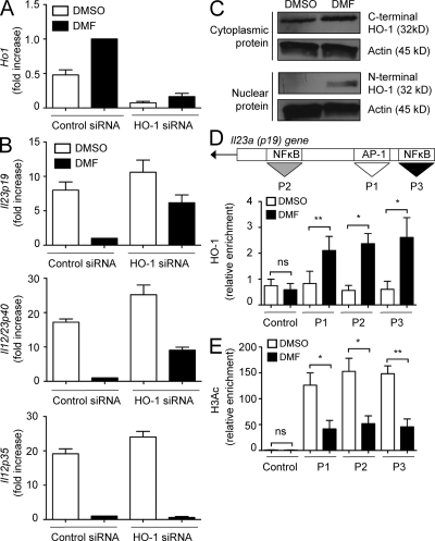
Figure 3.
DMF-induced HO-1 selectively prevents IL-23 induction. (A) DCs were treated with DMSO or 70 µM DMF, and HO-1 mRNA expression was determined by quantitative RT-PCR. HO-1 data were normalized to β-actin, and HO-1 level in control siRNA–transfected DMF–treated DCs was set as 1.0. The results are representative of three independent experiments. Error bars represent SEM. (B) HO-1 was knocked down, and levels of IL-12/IL-23p40, IL-23p19, or IL-12p35 mRNA were determined by RT-PCR. Data (mean ± SEM) were normalized to β-actin, and message levels in control siRNA–transfected DMF-treated DCs were set as 1.0. (C) DCs were treated as in A and lysed, and nuclear or cytoplasmic cell extracts were analyzed by Western blotting using antibodies directed against C- or N-terminal HO-1 protein. (D and E) DCs treated as in A were activated with LPS, cross-linked, and immunoprecipitated with anti–HO-1 (D) or anti-H3Ac (E). Bound DNA was amplified by quantitative PCR for primer sites P1 (AP-1; position 412–422 bp), P2 (c-Rel; position 560–584 bp), and P3 (RelA/c-Rel; position 394–406 bp). Data were pooled from four separate experiments and represent mean ± SEM (*, P < 0.05; **, P < 0.01; ns, not significant).

