Figure 7.
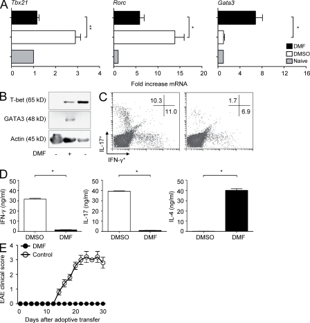
Type II DCs resulting from fumarate treatment selectively induce Th2 cells in vitro. (A) Naive OVA-specific CD4+ T cells were left unstimulated (gray bars) or were primed in vitro with OVA peptide and LPS-activated, DMSO-treated (open bars), or DMF-treated (black bars) DCs. Expression of the indicated transcription factors was assessed by RT-PCR, and the data were normalized to β-actin. Expression levels in unstimulated T cells were set at 1.0 (gray bars). Data are representative of three independent experiments (mean ± SEM; *, P < 0.05; **, P < 0.01). (B) DCs were treated and activated as in A and incubated alone (left lane) or with OVA-specific CD4+ T cells (middle and right lanes). T-bet or GATA3 protein expression in cell extracts was analyzed by Western blotting. (C and D) Naive OVA-specific CD4+ T cells were primed in vitro with OVA peptide and LPS-activated APCs in the presence or absence of DMF. Cells were expanded for 1 wk and then restimulated with OVA peptide and fresh APCs. Cytokines were determined by intracellular cytokine staining and flow cytometry (C) or by ELISA (D). Data from one representative experiment of three are shown (mean ± SEM; *, P < 0.001). (E) CD4+ T cells from immunized SJL mice were primed in vitro with PLP139-151 and APCs in the presence of DMF or DMSO (control) for 1 wk, restimulated, and expanded. 107 T cells were transferred into syngeneic WT mice (n = 5 per group), and EAE scores were determined. Data from one representative experiment of three are shown. Error bars represent SEM.

