Figure 8.
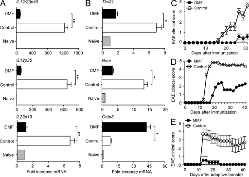
Fumarates induce type II APCs and Th2 cells in vivo and abolish the capacity of autoreactive CD4+ T cells to induce EAE. (A and B) SJL mice received DMF or DMF-free (control) water and were immunized with PLP139-151 peptide in CFA. On days 1–3, draining lymph nodes were isolated, RNA was extracted, and IL-12/IL-23p40, IL-12p35, or IL-23p19 expression (A) or Tbx21, Rorc, or Gata3 expression (B) was analyzed by RT-PCR (mean ± SEM; **, P < 0.001). Data were normalized to β-actin levels, and expression in naive mice was set as 1.0 (gray bars). Data from one representative experiment of three are shown (*, P < 0.01). (C) SJL mice were fed with DMF-containing or -free (control) water and immunized with PLP139-151 peptide in CFA and pertussis toxin (n = 5 per group). Clinical EAE scores were determined at the indicated times after immunization. Data are from one representative experiment of four. (D) TCR Vβ8.2 transgenic B10.PL mice were fed with 5 mg MMF or MMF-free (control) water (n = 8 per group). Mice were then immunized with MBP Ac1-11 peptide in CFA, and clinical scores were assessed at the indicated times after immunization. EAE incidence was 8/8 for control mice and 6/8 in the MMF group. (E) CD4+ T cells were isolated from spleens of MMF-treated or control donors from D on day 42, and equal numbers of cells (107) were adoptively transferred into naive mice. EAE scores were assessed at the indicated times after adoptive transfer (MMF group, n = 4; control group, n = 6). Experiments with MMF were performed four times in B10.PL or SJL mice, and data shown are from one representative experiment. (C and E) Error bars represent SEM.

