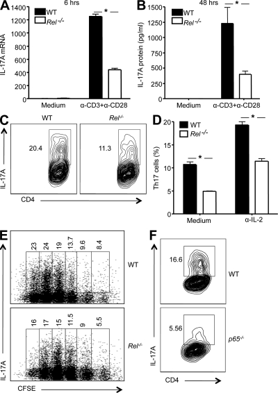
Figure 1.
T cells deficient in c-Rel or p65 are significantly compromised in their IL-17 gene expression and Th17 differentiation. (A and B) CD4+ T cells were isolated from 6-wk-old WT and Rel−/− mice (n = 5), and stimulated with anti-CD3 and anti-CD28 in the presence of anti–IL-2 and anti–IFN-γ for 6 h (A), or 48 h (B). Total RNA was extracted, and IL-17 mRNA levels were determined by real-time RT-PCR (A). The cytokine concentrations in the culture supernatants were determined by ELISA (B). (C and D) Purified CD4+ naive splenic T cells from WT and Rel−/− mice (n = 5) were cultured under Th17 cell–inducing conditions, as described in Materials and methods, with or without anti–IL-2 for 3 d. Cells were then restimulated with PMA and ionomycin in the presence of GolgiStop for 4 h, stained intracellularly with antibodies to IL-17A, and analyzed by flow cytometry. In C, IL-17A expression level of cells cultured with anti–IL-2 was plotted against CD4. In D, the percentages of IL-17A-expressing cells (as shown in C) from each group were compared. Error bars indicate the SDs of the means. (E) Purified CD4+ naive splenic T cells from WT and Rel−/− mice (n = 3) were incubated with 5 µM of CFSE for 10 min. After washing, cells were cultured under the Th17 cell–inducing condition, as described in Materials and methods. 3 d later, cells were restimulated with PMA and ionomycin in the presence of GolgiStop for 4 h, stained intracellularly with antibodies to IL-17A, and analyzed by flow cytometry. Each numerical figure in the graph represents the percentage of cells with the same number of divisions in the gated area. (F) p65−/− and p65+/+ chimeric mice were generated by adoptively transferring p65−/− and p65+/+ fetal liver cells, respectively, into irradiated B6 recipients as previously described (Ouaaz et al., 2002). 6 wk later, mice were sacrificed and CD4+ naive splenic T cells isolated. Cells were cultured under Th17 cell–inducing medium for 3 d, as described in Materials and methods. They were then restimulated and tested as described in C. The number in each panel represents the percentage of gated IL-17A–producing cells in total CD4+ cells. Results are representative of three independent experiments. *, P < 0.01.

