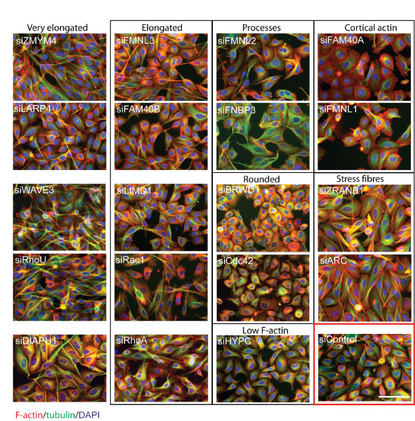
Figure 1.
Effects of PMM depletion on cell morphology. PC3 cells were transfected with the indicated siRNA pools for each PMM or for known regulators of actin dynamics in 384-well plates. After 72 h, cells were fixed and stained for F-actin (red), tubulin (green) and nuclei (blue). Cells are divided into groups based on their morphology. Scale bar, 100 μm.

