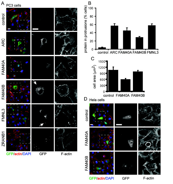
Figure 8.
PMM overexpression alters actin organization. PC3 cells (A) or HeLa cells (D) plated on Matrigel were transfected with constructs encoding the indicated N-terminal GFP-tagged PMM proteins (ARC, FAM40A, FAM40B, FMNL3 and ZRANB1) or GFP alone (control). After 18 h, cells were fixed and stained for F-actin, α-tubulin and DNA (DAPI). Scale bars, 10 μm. Boxed areas in merged images are enlarged to show detail of F-actin and PMM localization. Arrowheads (A) indicate FAM40B and FMNL3 localization to protrusions. (B) Quantification of PMM protein localization in protrusions in transfected PC3 cells. Control is GFP alone. Values are means ± S.D. of 26 to 66 cells per condition in each of three independent experiments. (C) Cell area (μm2) of transfected PC3 cells. Values are means ± s.e.m. of 50 to 100 cells in three independent experiments. ***P ≤ 0.001, control vs. FAM40A (unpaired Student's t-test).

