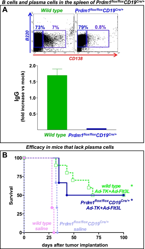
Figure 5.
Lack of plasma cells does not impair the efficacy of Ad-TK+Ad-Flt3L. (A) Representative dot plots show the percentage of B cells (B220+) and plasma cells (B220+CD138+) as assessed by flow cytometry. Note the lack of plasma cells in Prdm1flox/flox CD19Cre/+ mice. Splenocytes were collected from wild-type and Prdm1flox/floxCD19Cre/+ mice and incubated in the presence of 10 µg/ml LPS for 72 hours. Total IgG levels (IgG1, IgG2, IgG3, and IgG4) were assessed using an Easy-Titer IgG Assay Kit. (B) Kaplan-Meier curves show the survival of wild-type or Prdm1flox/floxCD19Cre/+ mice, which are unable to produce antibodies, that were implanted in the brain with GL26 cells and treated 14 days later with Ad-TK+Ad-Flt3L or saline. *P < .05 versus saline. Mantel log-rank test.

