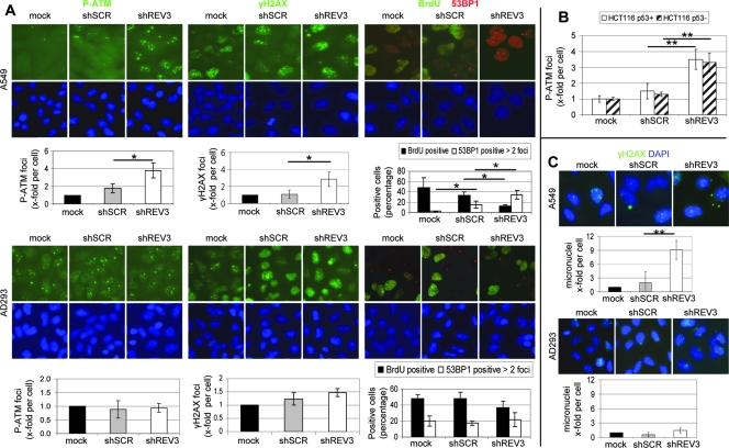
Figure 2.
REV3 depletion induces persistent DNA damage and genomic instability specifically in cancer cells. Cells were mock treated or transduced with lentiviral-based particles containing either shSCR or shREV3 and analyzed after 1 week. (A) Cells were stained for P-ATM, γH2AX, or BrdU (all green) and 53BP1 (red) and quantified by immunofluorescence microscopy. Cells containing more than two 53BP1 foci per cell were considered as 53BP1 positive. (B) Cells were stained for P-ATM, and foci per cell were quantified by immunofluorescence microscopy. (C) Cells were stained for γH2AX (green) and nuclear DNA was labeled with DAPI (blue). Micronuclei formation was identified by immunofluorescence microscopy-based analysis of DAPI staining. At least three independent experiments were analyzed. *P < .05. **P < .01. Shown are means ± SD.

