Fig. 5.
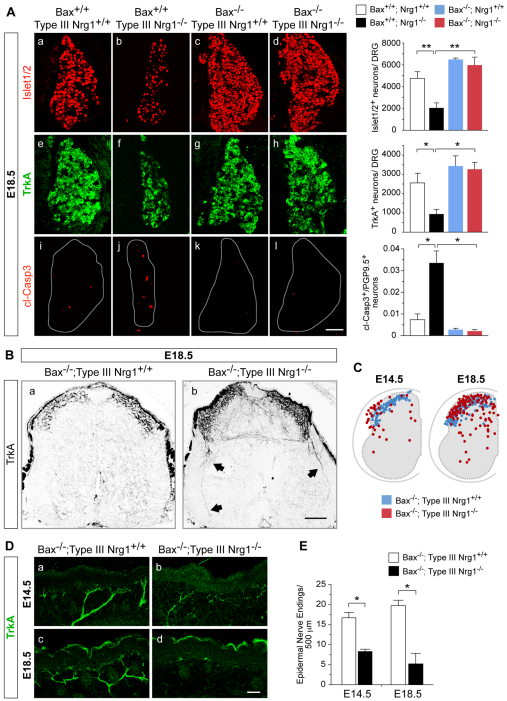
TrkA+ central projections and cutaneous innervation are aberrant in Bax–/–; type III Nrg1–/– mutants. (A) Sections of lumbar DRG from mouse embryos of the indicated genotypes at E18.5 were labeled for Islet1/2 (a-d), TrkA (e-h) or cl-Casp3 (i-l). Bar charts show quantification of Islet1/2+, TrkA+ and cl-Casp3+ neurons in DRG at E18.5. (B) Spinal cord sections from Bax–/–; type III Nrg1+/+ (a) or Bax–/–; type III Nrg1–/– embryos (b) at E18.5 were labeled for TrkA. Aberrant TrkA+ axons were detected in the double-null spinal cord at E18.5 (arrows). (C) Summary plots of individual TrkA+ axon termination points within Bax–/–; type III Nrg1+/+ (blue dots) or Bax–/–; type III Nrg1–/– (red dots) spinal cords at E14.5 and E18.5 (n=3 spinal cords per genotype). (D) Sections of hindlimb epithelium from Bax–/–; type III Nrg1+/+ (a,c) or Bax–/–; type III Nrg1–/– (b,d) embryos at E14.5 or E18.5 were labeled for TrkA. In double-mutant embryos, TrkA+ axons appeared thin and disorganized at E14.5 (b), and by E18.5 (d) axon innervation was reduced. (E) Quantification of TrkA+ nerve endings in hindlimb epithelium (n≥2 embryos per genotype). Mean ± s.e.m. *, P<0.02; **, P<0.005 (ANOVA with post-hoc Fisher’s PLSD test). Scale bars: 100 μm in A,B; 50 μm in D.

