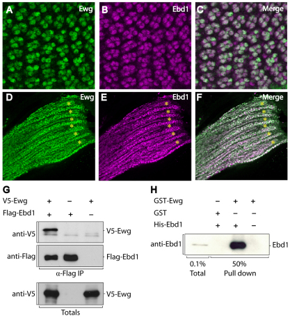
Fig. 4.
Ewg and Ebd1 display similar expression patterns and interact directly. (A-F) Confocal images of pupal retinas at 24 hours after puparium formation (A-C), pupal myofibers and associated myoblasts (D-F). Tissues are immunostained with Ewg antiserum (A,D) and Ebd1 antiserum (B,E). Merged images are shown in C,F. (A-F) During metamorphosis, Ewg and Ebd1 colocalize in neurons and myocytes, including photoreceptors (A-C), developing myofibers (asterisks) and associated myoblasts (D-F). (G) V5-tagged Ewg and Flag-tagged Ebd1 were co-transfected into HEK 293T cells. Cell lysates were subjected to immunoprecipiation with anti-Flag antibody, followed by immunoblotting with anti-V5 and anti-Flag antibody. Immunoblotting of total lysates is shown in lower panel. (H) GST interaction assay. Bacterially expressed GST-Ewg or GST control were purified and incubated with bacterially expressed and purified His-Ebd1. Protein bound to GST-Ewg or GST control was detected by immunoblotting with Ebd1 antibody.

