
Figure 7.
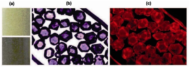
Optical observation of the raw 80–100 mesh HPHT diamond. (a) HPHT microdiamond seen with the naked eye: initial (above) and after electron irradiation (below), (b) Bright-field transmission image of the microdiamond after electron irradiation and annealing. (c) Image of the microdiamond after electron irradiation and annealing recorded with a fluorescence microscope under green excitation (20 ms duration).