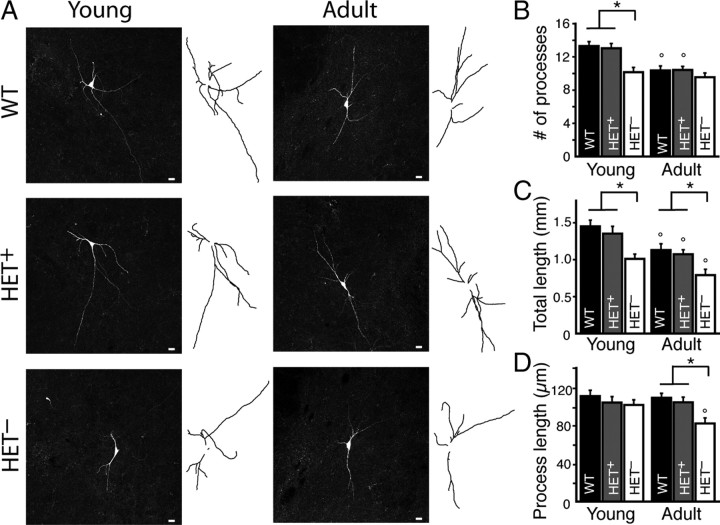
Figure 2.
Mecp2− dopamine neurons in Mecp2+/− mice (HET−) have a reduced dendritic arbor compared to Mecp2+ neurons from WT or Mecp2+/− (HET+) female mice. A, Representative maximum intensity projection confocal images and tracings of neurobiotin-filled WT, HET+, and HET− dopamine neurons from the SN of young and adult wild-type and Mecp2+/− mice. B, The number of processes (dendrites and axon) on dopamine neurons decreased with age in wild-type and Mecp2+/− mice, indicating a developmental pruning of dendrites. HET− neurons had fewer dendrites than WT and HET+ neurons in young animals. There was no difference in the number of dendrites on WT and HET+ neurons. C, The total length of processes (in mm) decreased with age in wild-type and Mecp2+/− mice. The total length of processes in HET− neurons was significantly less than those in WT or HET+ neurons in both young and adult animals. There was no difference in the total length of processes in WT and HET+ neurons. D, The average process length (in μm) of WT and HET+ neurons was not different and did not change with age. Processes from HET− neurons from adult Mecp2+/− mice were significantly shorter than age-matched WT and HET+ neurons. Mean ± SEM; asterisk (*) designates statistical significance within age group; circle (°) designates statistical significance compared to young age.

