
Figure 6.
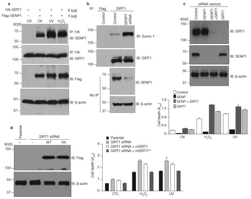
SENP1 interacts with and desumoylates SIRT1 and promotes stress-induced apoptosis. (a) Cells were cotransfected with HA–SIRT1 and Flag–SENP1 and treated with UV radiation or hydrogen peroxide. HA immunoprecipitates were immunoblotted with antibodies against SENP1 or SIRT1. Cell extracts were immunoblotted with antibody against Flag or β-actin. (b) SENP1 siRNA was stably transfected into H1299 cells. Endogenous SENP1 expression, and SIRT1 expression and sumoylation, were determined by immunoblotting. (c) H1299 cells stably expressing control vector, SENP1 siRNA, SIRT1 siRNA or both were treated with UV radiation or hydrogen peroxide. Amounts of the indicated proteins were determined by western blotting. Cell apoptosis was determined by measuring DNA fragmentation by ELISA. Each data point is the average of triplicate samples (n = 3) analysed in parallel. The error bars represent s.d. (d) H1299 cells stably expressing SIRT1 siRNA were transfected with control plasmid or expression vectors for Flag-tagged wild-type or mutant mouse SIRT1 (mSIRT1). The expression of mouse SIRT1 and β-actin was determined by immunolotting. Cellular apoptosis was determined by measuring DNA fragmentation by ELISA. Each data point is the average of triplicate samples (n = 3) analysed in parallel. The error bars represent s.d. Uncropped images of the scans are shown in the Supplementary Information, Fig. S1.