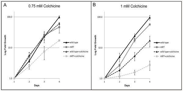
Figure 7.
mlt1 cells exhibit enhanced sensitivity to colchicine. Graphs plotting the average logarithmic-fold concentrations of triplicate samples of wild-type and mlt1 cells ± standard deviation grown with and without colchicine at concentrations of 0.75 and 1 mM. Data points were normalized to the first 24-hour counts. (A) With treatment at 0.75 mM colchicine, only mlt1 showed a significant retardation in growth at all time points (p value < 0.05). At day 4 both wild-type and mlt1 strains showed significant growth retardation with drug treatment (p value < 0.05), with wild-type doubling time reduced by one hour and mlt1 doubling time reduced by four hours relative to the non-treated controls. (B) At 1 mM colchicine, both strains showed a significant retardation of growth at all time points (p value < 0.05). At day 4 wild-type doubling time was reduced by six hours, whereas mlt1 doubling time was reduced by 38 hours.

