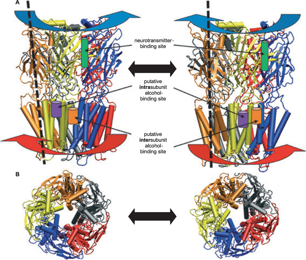
Fig. 3.
Models of 2 states of a ligand-gated ion channel with pentamer subunits colored individually (orange, gray, red, blue, yellow) illustrating “wringing” or iris-like gating motions. (A) Lateral view from the plane of the membrane. The ligand-binding extracellular (EC) domain (blue arrows) and transmembrane (TM) domain (red arrows) twist in opposite directions. The dotted lines illustrate tilted axes of spiral motion of 1 subunit in each state. These motions create a harmonic spiral motion that results in channel gating. One of 5 native agonist ligand-binding sites in the EC domain is illustrated as a cylinder (green) between 2 subunits. Putative binding sites for alcohols and anesthetics are illustrated as cylinders at intrasubunit (purple) and intersubunit (orange) helix interfaces within the TM domain. Note that, although only 1 example of each binding site is shown, the 5-fold symmetry or pseudosymmetry of the channel may create as many as 5 such sites either within or between subunits. (B) Top view of the ion pore from the EC side. Figure modified from Bertaccini and colleagues (2010a).

