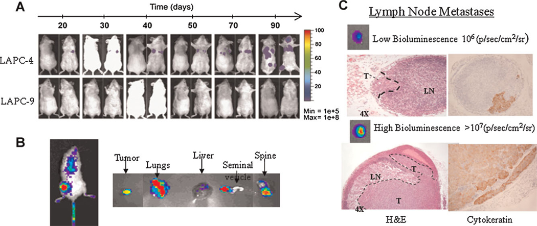
Fig. 3.
Bioluminescence imaging (BLI) to study experimental metastasis. A: The use of BLI to assess metastatic potentials of two prostate xenograft models (LAPC-4 and -9), longitudinal over time. The ease of use and low background of luciferase reporter genes are clear advantages.
B: BLI provides a 2-D semi-quantitative signal, which is difficult to localize in a metastatic setting. Imaging of harvested organs ex vivo can aid in analyze the magnitude and localization of tumor spread. C: The magnitude of bioluminescence signals in isolated organs correlates well with volume of metastasis, as assayed in the lymph node metastasis. These results are adapted from Brakenhielm et al. [78]. [Color figure can be viewed in the online issue, available at wileyonlinelibrary.com.]

