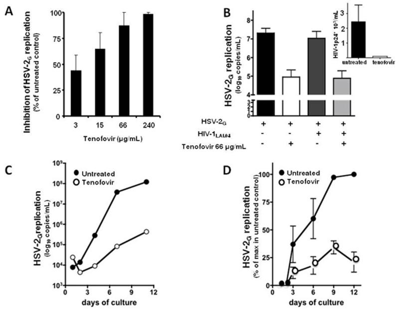
Figure 4.
Suppression of HSV-2 in singly infected and in HIV-1-coinfected human ex vivo lymphoid tissue by tenofovir.
a: Blocks of human tonsillar tissue were inoculated ex vivo with HSV-2G and treated or not with tenofovir (3, 15, 66, 240 μg/ml). HSV-2G replication was monitored by measuring viral DNA in culture media (day 9 post-infection). Presented are means ± SEM of the results with tissues from 14 donors. For each donor, each data point represents pooled viral release from 27 tissue blocks.
b: Blocks of human tonsillar tissue were co-inoculated ex vivo with HSV-2G and HIV-1LAI and treated or not with tenofovir (66 μg/ml). HIV-1 replication was monitored by measuring p24gag accumulated in culture media over 3 day periods. Presented are means ± SEM of the results with tissues from 6 donors. For each donor, each data point represents pooled viral release from 27 tissue blocks.
c: Blocks of human cervico-vaginal tissues were inoculated ex vivo with HSV-2G and treated or not with tenofovir (150 μg/ml). HSV-2G replication was monitored by measuring viral DNA accumulated in culture media at different time points throughout the culture period. Presented is a representative experiment (out of five) with a tissue from individual donor. Each data point represents pooled viral release from 16 tissue blocks.
d: Blocks of human cervico-vaginal tissues were inoculated ex vivo with HSV-2G and treated or not with tenofovir (150 μg/ml). HSV-2G replication was monitored by measuring viral DNA accumulated in culture media at different time-points throughout the culture period. Presented are means ± SEM of the results with tissues from 5 donors. For each donor, each data point represents pooled viral release from 16 tissue blocks.

