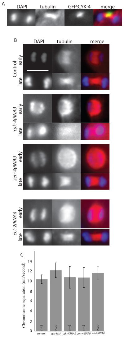
Figure 5. Meiotic spindle microtubule structure is unaffected by loss of centralspindlin.
(A) shows central spindle localization of GFP:CYK-4 on a fixed meiotic anaphase spindle, stained to show tubulin (red) and chromosomes (DAPI, blue). (B) shows meiotic anaphase spindles fixed and stained to show tubulin (red) and chromosomes (DAPI, blue) in early and late anaphase under conditions of control RNAi, cyk-4(RNAi), zen-4(RNAi) or ect-2(RNAi), as labeled. Scale bar, 5 μm. (C) shows anaphase chromosome separation rates for control or centralspindlin-depleted embryos (all, p > 0.1).

