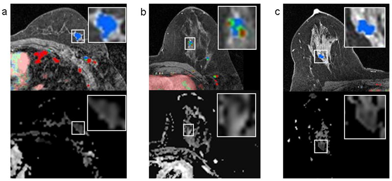
Figure 3.

Several lesion examples where DCE-MRI and DWI suggested different levels of suspicion. DCE-MR images are shown on the top row and corresponding ADC maps are shown below. In each case, the lesion of interest is delineated by a box on both images and enlarged for clarity (boxes do not represent the ROIs, which were manually drawn within lesion borders as described in the methods). A 1.1 cm invasive ductal carcinoma in a 59 year-old woman that demonstrated only persistent enhancement on DCE-MRI with no washout, but exhibited a low ADC value on DWI (mean ADC, 1.34 ×10⁻³ mm2/s) (a). A 1.0 cm benign papilloma detected in a 41 year-old woman that demonstrated concerning washout (red), but exhibited high ADC (mean ADC, 2.04 ×10⁻³ mm2/s) (b). A 2.6 cm region of benign fibrocystic change detected in a 43-year old woman that demonstrated no washout on DCE-MRI, but exhibited suspiciously reduced diffusivity, with low ADC values (mean, 1.04 ×10⁻³ mm2/s).
