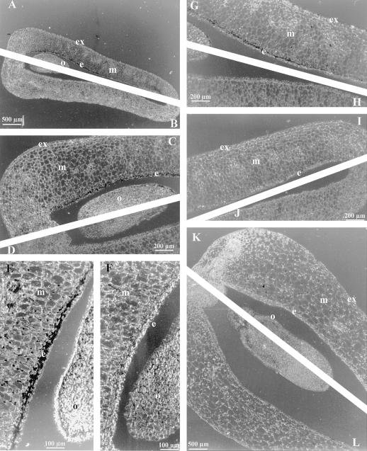
Figure 5.
Detection of tissue-specific TPE4A expression by in situ hybridization. A through J, Cross-sections of senescent pea ovaries (d 4 after anthesis) hybridized with the TPE4A antisense riboprobe (A, C, E, G, and I) and the sense control probe (B, D, F, H, and J). C and D, Ovule and surrounding area of the sections shown in A and B at higher magnification; G, H, I, and J, other parts of the same sections. E and F, Detail of the ovule and surrounding endocarp area of different sections. K and L, Cross-section of the ovule and surrounding endocarp area of developing pea fruits (GA3-treated ovaries on d 4 after anthesis) hybridized with the TPE4A antisense riboprobe (K) and the sense control probe (L). Hybridization is indicated by the dark staining in the endocarp (e) area surrounding the ovule (o) and, at lower levels, in the outer cell layers of the ovule in senescent ovaries. m, Mesocarp; ex, exocarp.

