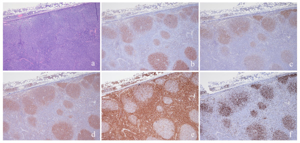
Figure 2.
Invasive follicular lymphoma. FL is formed by large nodules with interfollicular invasion (a) showing positive staining for CD10 (b), BCL6 (c), HGAL (d); the positivity for GC markers are more evident in the nodular component than in the interfollicular areas in this case. BCL2 was moderately and partially expressed (e). Ki67 proliferative index was high (f).

