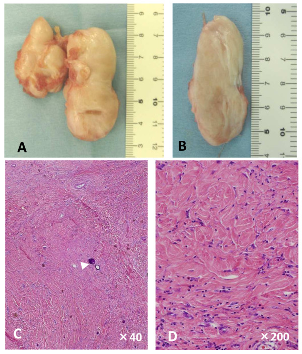
Figure 2.
Macroscopic (A and B) and microscopic (C and D) features of the resected mass. The two lesions were found to be solid, lobular, well-circumscribed, and gray-white masses. (A) The two nodules were connected with each other by fibrous tissue. (B) The cut surface was gray-white and homogenous. (C, D) Hematoxylin and eosin staining of the lesions revealed that they were composed of hyalinized collagen fibers with fibroblasts. Psammomatous calcifications were observed (C, arrowhead). Original magnifications: (C) ×40; (D) ×200.

